Bridging the gap between structural and functional vision problems.
Traditional eyecare providers focus on diagnosing and maintaining ocular health. The eyes are literally the windows to the brain and body. The early signs of high blood pressure, diabetes, and a range of systemic diseases can be detected through an eye health exam.
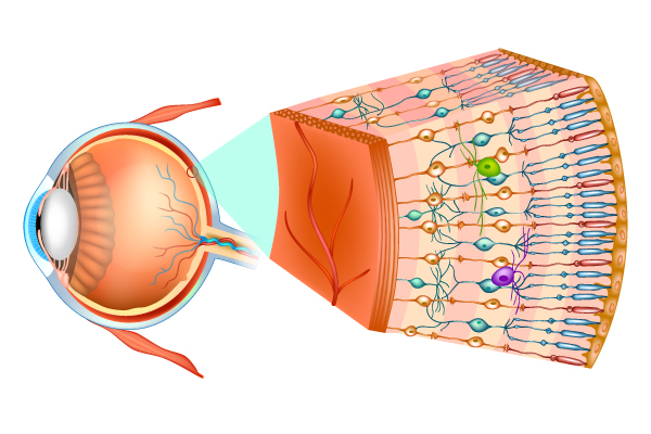
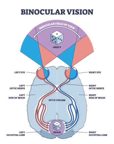
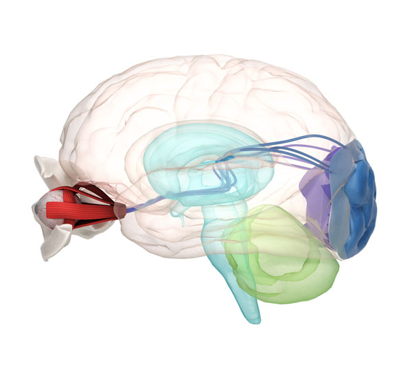
To see well, the tissues that bend the light at the front and the back of the eye (retina) have to be healthy. Visual information then travels as electrical impulses from the retina back through the optic nerve to the visual cortex at the back of the brain. Beyond this point, visual information is sent to various parts of the brain for visual perception, visual memory, motor planning and execution.
At Ho Vision Group, we have access to state-of-the-art technology to detect damage to the eye and brain structures related to visual processing by:
A retina camera and optic coherence tomography (OCT) that can detect damage to the retina and optic nerve (brain tissues) earlier than traditional eye exams. This allows for early intervention, counselling, and careful monitoring of disease progression for macular degeneration and glaucoma for example.
Automated visual fields (VF) that can detect central and peripheral fields loss resulting from damage to the pathway extending from the retina and optic nerve to the visual cortex in the brain. This allows for monitoring of disease progression of eyeball structures and characterizing damage to brain pathways in stroke and brain injury. Fields are also a test for whether patients have enough peripheral fields to drive.
Visual evoked potential (VEP) that can detect electric signal imbalances in central or peripheral visual information and between the right and left visual pathway (extending from retina to the brain). This is helpful for determining the degree of damage and prognosis in open head trauma cases.
Electroretinogram (ERG) that can detect electric signal decreases from information transmitted by structures such as optic nerve or retina.
The functional aspect of the visual exam assesses: visual acuity, eye alignment, eye teaming, eye focusing, visual stamina, and visual integration with other sensory-motor systems in the body. Many patients with concussions may have 20/20 eyesight and a healthy eyeball yet fail this part of the assessment.
All this information comes together to provide our doctor with the best knowledge on the stability of a visual problem - its structural and functional deficits. This gives our doctor a realistic expectation on the aids that can be used and the extent of improvement for visual rehabilitation.
At Ho Vision Group, we take an individualized approach to every patient to tailor a product or program to help patients achieve their goals. We also recognize that success is also driven by patients’ personal.
Define the Vision Problem
Eye health
Functional
or Both
Treatment Plan
Lenses
Tools
Vision rehabilitation
Improve Vision Function and Improve Quality of Life