-
Low vision can be a challenging and sometimes confusing condition, especially when it comes to understanding how it impacts the way our eyes work together. To grasp this better, let’s start with a basic concept: how our eyes normally work together to give us a clear, single view of the world.
How Our Eyes Work Together: Bifoveal Fixation
In a healthy vision system, both eyes work in tandem to focus on a single point. This process is called bifoveal fixation. Here’s a simplified breakdown:
- Fovea: Each eye has a small, central part of the retina called the macula and within this area lies the fovea. This is where our vision is sharpest. This central area also has the most cones, these are cells responsible for color vision.
- Fixation: When we look at something, the fovea of both eyes align so that the image falls directly on the fovea of each eye. If the fovea of one eye is misaligned, a person can see double vision or may complain of visual blur.
- Brain Processing: The brain receives these sharp images from both eyes and merges them into a single, clear picture. This precise alignment also helps our brain judge distances and depth accurately.
The Role of Macula in Vision
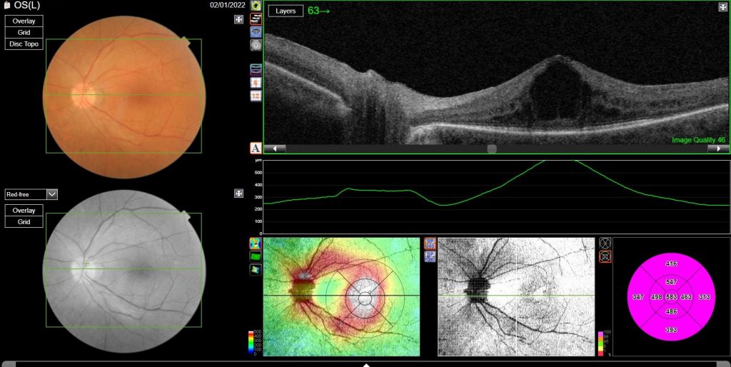
The macula is a crucial part of the retina that includes the fovea. It plays a key role in central vision, which we use for activities like reading, driving, and recognizing faces. When the macula is healthy, it ensures that the images falling on the fovea are sharp and detailed. Many low vision patients struggle with blur, visual distortion, and/or light sensitivity due to damage from the retina. The imperfect image is similar to developing a picture from a damaged computer chip or film.
Impact of Low Vision on Eye Teaming
Eye diseases like macular degeneration or diabetic retinopathy can result in an unhealthy macula. When the macula is damaged, it changes how the eyes work together. Here’s how:
- Parafoveal Fixation: With macular problems, the brain may use areas of retina that are less damage around the damaged fovea. These regions are less precise, leading to a loss of sharpness for the image that is already degraded by the physical damage from the retina.
- Misalignment: Because the parafoveal areas are not as finely tuned as the fovea, the eyes may struggle to align perfectly. In children, strabismus (or an eye turn) can occur when the brain chooses to not use the fovea to fixate. In low vision, strabismus can be a strategy the brain adapts to due to damage of the fovea. In either case, misalignment can cause blur and/or double vision.
It gets even more complicated when the damage is not the same in both eyes causing asymmetrical distortion. Some of Dr. Ho’s patients have complained about the image in one eye being smaller or more crooked from the other. - Impaired Spatial Judgment: Since accurate spatial judgment relies on the fovea aligning from both eyes, parafoveal fixation can result in difficulties in judging distances and depth. This can affect daily activities, making tasks like walking, picking up objects, or even pouring a drink more challenging.
Why Understanding Brain Strategy Matters
Understanding how the brain changes strategy in eye teaming for low vision sets the stage for doctors to use different tools to help improve their patient’s quality of life. It may not be as simple as providing appropriate magnification. Glasses can be outfitted with many types of support (prism, tint, optical magnification, selective blur) to optimize outcome. Dr. Ho also introduced using optometric syntonic therapy to help the brain (cortical) transition to a less disruptive adaptation.
If you are interested in listening to the full lecture, feel free to download Dr. Ho’s lecture from Optometric Extension Program Foundation (OEPF) here:
Strategies on Imperfect Fusion-Binocular considerations for Low Vision

